
D. I. Amarasinghe, Department of Radiography & Radiotherapy, Faculty of Allied Health Sciences, General Sir John Kotelawala Defence University
Cancer is one of the most lethal diseases globally due to its aggressive and heterogeneous nature. It often begins as a small cluster of mutated cells within complex tissue environments, making early diagnosis challenging. Imagine if a single Magnetic Resonance Imaging (MRI) could predict how a cancer might behave, guiding treatment decisions before a biopsy is performed, this is the promise of radiomics.
What is Radiomics?
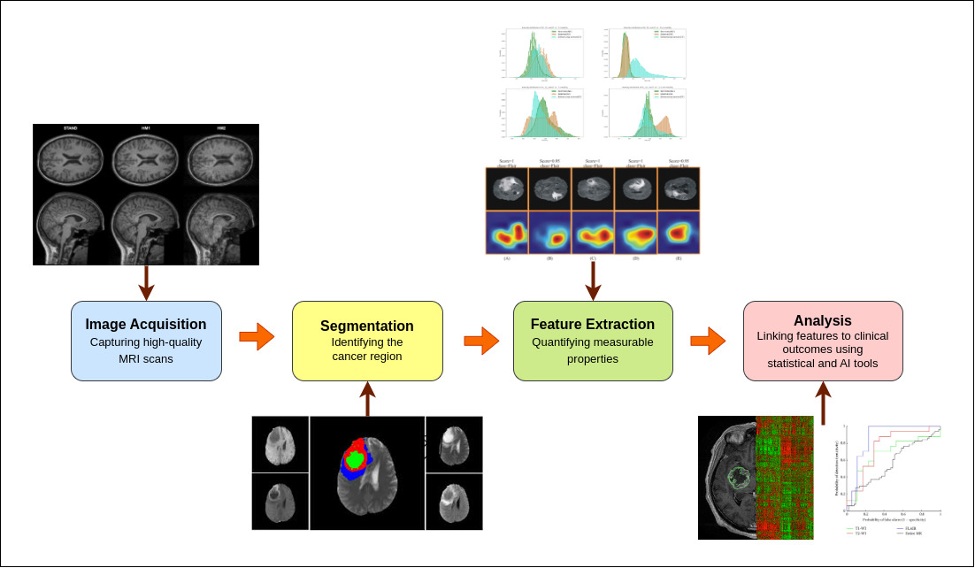
Radiomics is an emerging field that uses advanced computational methods to extract quantitative data from medical images, uncovering patterns invisible to the human eye. Since its introduction in 2012, radiomics has gained momentum due to advancements in imaging technology and computational power, enabling more complex analyses of medical data [1].
By quantifying structural information such as shape, texture, and intensity within cancers, radiomics provides predictive insights into treatment outcomes and genetic profiles. This approach is similar to texture analysis in agriculture, which assesses the quality of soil or harvests, as it characterizes the “texture” of cancers to enhance understanding of their properties.
Why MRI is Ideal for Radiomics?
MRI is particularly suited for radiomics due to its high soft tissue differentiation, allowing clear visualization of cancers and their surroundings. Unlike other imaging modalities, MRI uses no radiation, ensuring patient safety during repeat scans required for ongoing monitoring. Its multi-parametric imaging capabilities, such as T2-weighted imaging (T2WI), apparent diffusion coefficient (ADC) maps, and multi-phase contrast-enhanced sequences, provide a comprehensive view of cancer features.
For example, MRI-based radiomic features can differentiate between high-grade and low-grade cancers in Glioblastoma, offering critical diagnostic insights [2]. These enhanced imaging features facilitate extracting a wide range of quantitative data, improving cancer characterization and diagnosis in clinical practice.
Predictive Biomarkers in Cancer Treatment
Predictive biomarkers are vital in cancer treatment as they provide measurable characteristics that guide therapeutic decisions based on cancer behavior. MRI-based biomarkers identify normal biological processes, pathogenetic changes, and therapeutic responses. As examples,
- In glioblastoma, MRI features can predict the Isocitrate Dehydrogenase (IDH) mutation status, a crucial factor in the diagnosis, prognosis, and treatment of gliomas [3].
- In breast cancer, radiomics can predict responses to neoadjuvant chemotherapy [4].
- In non-small cell lung cancer, radiomic biomarkers achieved 87.42% accuracy in predicting outcomes for patients undergoing immunotherapy [5].
Challenges and Future Directions
Despite its potential, the broad use of MRI-based radiomics faces several challenges. One of the major issues is the lack of standardization in pre-operative MRI protocols, complicating result comparisons and model validation. Variations in MRI scanner settings can lead to inconsistent radiomic feature values, impacting reproducibility. Furthermore, analyzing high dimensional radiomic data requires advanced computational tools and regulatory hurdles must be addressed for successful clinical implementation [6].
Looking forward, the future of MRI-based radiomics is promising. The research will focus on incorporating diverse imaging sequences to extract more informative features and improve model stability [7]. Advancements in data standardization, interpretability of deep learning models, and the development of user-friendly predictive tools will be essential for clinical acceptance and effective implementation in oncology practice. As ongoing research addresses current challenges, MRI-based radiomics has immense potential as a predictive biomarker in cancer, to revolutionize cancer management and improve patient outcomes.
References
[1] W. Rogers et al., “Radiomics: from qualitative to quantitative imaging,” The British Journal of Radiology, vol. 93, no. 1108, p. 20190948, Apr. 2020, doi: https://doi.org/10.1259/bjr.20190948.
[2] G. W. Hooper and D. T. Ginat, “MRI radiomics and potential applications to glioblastoma,” Frontiers in Oncology, vol. 13, Feb. 2023, doi: https://doi.org/10.3389/fonc.2023.1134109.
[3] H. Ding et al., “Prediction of IDH Status Through MRI Features and Enlightened Reflection on the Delineation of Target Volume in Low-Grade Gliomas,” Technology in Cancer Research & Treatment, vol. 18, p. 153303381987716-153303381987716, Jan. 2019, doi: https://doi.org/10.1177/1533033819877167.
[4] P. McAnena et al., “A radiomic model to classify response to neoadjuvant chemotherapy in breast cancer,” BMC Medical Imaging, vol. 22, no. 1, Dec. 2022, doi: https://doi.org/10.1186/s12880-022-00956-6.
[5] J. Peng, D. Zou, X. Zhang, H. Ma, L. Han, and B. Yao, “A novel sub-regional radiomics model to predict immunotherapy response in non-small cell lung carcinoma,” Journal of Translational Medicine, vol. 22, no. 1, Jan. 2024, doi: https://doi.org/10.1186/s12967-024-04904-6.
[6] J. E. van Timmeren, D. Cester, S. Tanadini-Lang, H. Alkadhi, and B. Baessler, “Radiomics in medical imaging—‘how-to’ guide and critical reflection,” Insights into Imaging, vol. 11, no. 1, Aug. 2020, doi: https://doi.org/10.1186/s13244-020-00887-2.
[7] J. Bao et al., “Prediction of clinically significant prostate cancer using radiomics models in real-world clinical practice: a retrospective multicenter study,” Insights into imaging, vol. 15, no. 1, Feb. 2024, doi: https://doi.org/10.1186/s13244-024-01631-w.